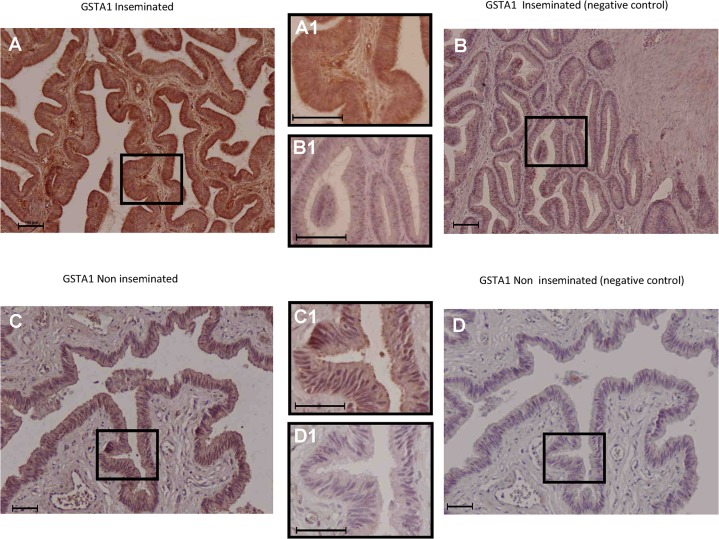
Fig 6. Immunohistochemical localization of GSTA1 in the porcine oviduct.
GSTA1 protein expression extended from the epithelial cells to deeper layers of the oviductal wall in the inseminated sows (Fig 6A, magnified in 6A1) compared with the non-inseminated animals, where labelling was mainly observed at the epithelial level (Fig 6C, magnified in 6C1). The corresponding negative controls are shown in Fig 6B, magnified in 6B1 for the inseminated animals and in Fig 6D, magnified in 6D1, for the non-inseminated sow. In all figures, scale bars correspond to 100 μm.

