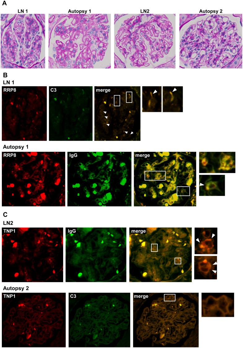
Fig 2. Double immunofluorescence of RRP8 or TNP1 and IgG or C3 in renal sections from patients with LN.
Formalin-fixed, paraffin-embedded sections of biopsy or autopsy specimens from LN patients were processed for double immunofluorescence staining. Representative photographs are shown. The specimens were stained with Alexa 546-conjugated anti-RRP8 or anti-TNP1 antibodies (red) and with FITC-conjugated anti-IgG or anti-C3 antibodies (green). (A) The same glomeruli were stained with periodic acid-Schiff. (B) Co-presence of RRP8 and C3 was detected along the sub-epithelial area of the glomeruli showing a dotted pattern (arrowhead) in LN1. Also in Autopsy1, both RRP8 and IgG signals were detected in the mesangial area as well as in the sub-epithelial area (arrowhead). (C) Co-presence of TNP1 and IgG was revealed along the basement membrane showing a linear pattern in LN2, as was the case for TNP1 and C3 in Autopsy2. The bold and strong autofluorescence in each of the panels is mainly due to red blood cells.

