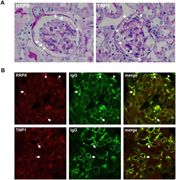
Fig 7. Double immunofluorescence of RRP8 or TNP1 and IgG in renal sections from RRP8-injected or TNP1-injected mice.
Cryostat kidney sections from mice were analyzed by double immunofluorescence staining with a combination of anti-human RRP8 or anti-human TNP1 antibody and anti-mouse IgG antibody. The specimens were stained with Alexa 568-conjugated anti-RRP8 or anti-TNP1 antibodies (red) and with Alexa 488-conjugated anti-murine IgG antibody (green). (A) Photomicrograph of representative glomeruli of RRP8- and TNP1-injected mice (periodic acid-Schiff staining). Both manifested the slight enlargement of glomeruli with thickening of capillary wall (arrows) as well as the endocapillary proliferation with leukocytes (arrow head). (B) RRP8 signals coexisted with IgG signals in the sub-endothelial (arrows) and sub-epithelial (arrowhead) regions, exhibiting a granular pattern. On the other hand, TNP1 signals coexisted with IgG signals in the sub-epithelial region (arrows).

