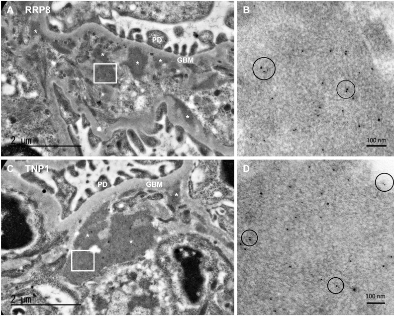
Fig 8. Immunoelectron microscopy of kidney sections from RRP8-injected (A and B) or TNP1-injected (C and D) mice.
Photographs show the characteristic dense deposit in the subendothelial part especially at around the border of pericapillary and perimesangial areas. Panel B (anti-RRP8) and Panel D (anti-TNP1) show the high-power view of the deposition which manifests the adjacent (within 30nm) of RRP8 or TNP1 represented by 10-nm-gold particles and IgG represented by 5-nm-gold particles (circle area). *, electron-dense deposit; PD, podocyte; GBM, glomerular basement membrane.

