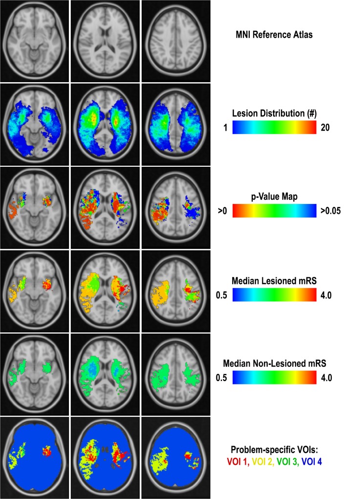
Fig 1. Illustration of the single processing steps used for generation of the problem-specific brain regions in three selected slices.
From top to bottom: MNI reference atlas, infarct distribution map used to exclude voxels lesioned in less than five patients from statistical calculations, p-value map used to exclude voxel with a significance level p≥0.05 from the VOI generation, median mRS values of lesioned and non-lesioned voxels used to define the final VOIs based on the median mRS difference d (VOI1: d > 2, VOI2: 1 < d ≤ 2, VOI3: 0 < d ≤ 1, VOI4: remaining voxels).

