
Fig 5.
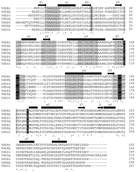
clustal omega alignment of known uronate dehydrogenases (Sievers et al., 2011). The secondary structures are shown above with thick bars for α-helices and arrows for β-sheets. Coloured residues represent conserved sequence motifs within the SDR family and also residues involved in the specific substrate and cofactor recognition.