
Fig 7.
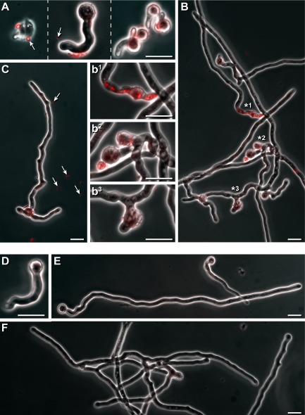
Effect of vesicle-containing droplets on A. proliferans.A–C. Conidia of A. proliferans were incubated 4 h in liquid medium. Then, an aliquot (20 μl) of vesicles containing droplets (see Fig. A and C) was added to a culture aliquot (500 μl), and incubation continued for additional 3 h incubated for 2 h at 30°C. (A) or for 8 h (B, C). All samples were inspected microscopically by phase contrast as well as for undecylprodigiosin-derived red florescence, and are shown in a merged fashion. Vesicle-like particles that are red fluorescent ( ) are marked. Regions from the hyphae (B) with high red intracellular fluorescence (*1, or that had developed blown up (*2), or strongly deformed (*3) areas are presented in an enlarged fashion (b1, b2 and b3). Bars 10 μm.D–F. Controls were without the addition of droplets, and inspected as outlined above. Bars 10 μm.