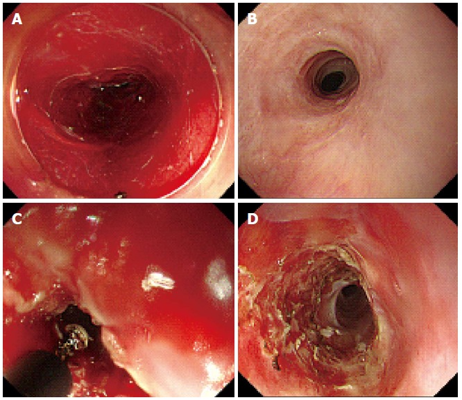
Figure 3.

Representative endoscopic images of refractory strictures of the esophagus. A: Endoscopic images immediately after 10-cm long circumferential ESD; B: Images 3 mo after ESD followed by repeated EBD with steroid injection; C: ERIC procedure with IT-2 knife (Olympus, Tokyo, Japan); D: Severe stricture 1 mo after ERIC. ESD: Endoscopic submucosal dissection; ERIC: Endoscopic radial incision and cutting; EBD: Endoscopic balloon dilation.
