
Figure 3.
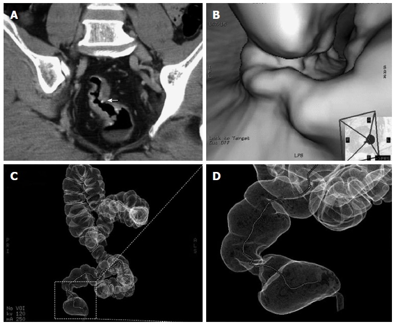
Computed tomography presentation. A: Computed tomography (CT) scan showing curvilinear calcification in the rectosigmoid colon and calcified, conglomerate nodules (arrow) protruding from the wall of the rectosigmoid colon; B: Lobulated polypus in the rectum; C, D: CTVC enables three-dimensional view of walls of the colon as a result of reconstruction of multislice CT images. The colorectal stenosis is showed in the area surrounding by dotted lines. CTVC: CT virtual colonoscopy.