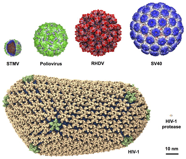
Figure 2.
Viral particles of different sizes studied using MD simulations. The viruses were arranged in the order of increasing size with the capsid diameters given in parentheses : STMV (17 nm) [20], poliovirus (32 nm) [22], RHDV (43 nm) [15], SV40 (49 nm) [23], and HIV-1 (70–100 nm) [1]. For size comparison, HIV-1 protease, one of the most studied enzymes, is shown at the bottom right.

