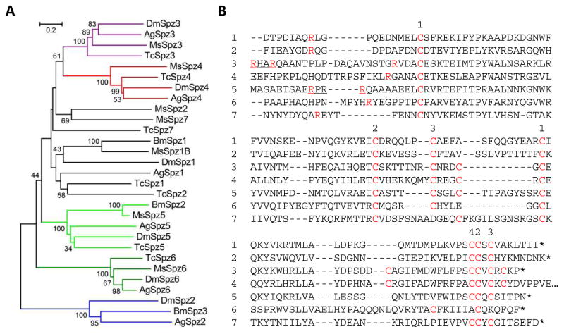
Fig. 1.
Phylogenetic relationships of Spätzles in M. sexta, B. mori, T. castaneum, and D. melanogaster. (A) Tree. Based on the sequence alignment of 29 full-length Spätzles, a tree was generated with branches shown in colors representing closely related groups. (B) Aligned sequences of the cystine-knot cytokine domains in M. sexta Spätzles-1 through 7. Cys residues are indicated in a red font. Some Cys residues may form intra- (1-1, 2-2, 3-3) and inter- (4) chain disulfide bonds. Proteolytic activation sites, known for Spätzle-1, are predicted to be next to the Arg (red) in Spätzle-2 through 6. The putative processing site (RXXR) is underlined in Spätzle-3 and 5.

