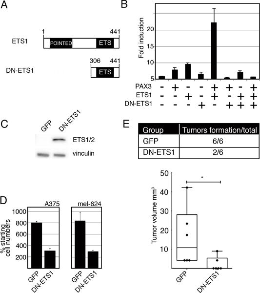
Figure 7.

Expression of a dominant-negative ETS1 (DN-ETS1) protein inhibits MET induction and melanoma cell growth. (A) A schematic of wild-type ETS1 and DN-ETS1 proteins. (B) DN-ETS1 inhibits the activation of the MET promoter by both PAX3 and ETS1. Luciferase assays of 293T cells transfected with METpm, in the presence (+) or absence (−) of PAX3, ETS1, and/or DN-ETS1 expression vectors. Fold induction was calculated by measuring luciferase activity in arbitrary light units, normalized against beta-galactosidase activity, and divided by the measurements obtained for reporter vector alone (n=9, p<0.005). (C) A western analysis detects DN-ETS1 expression in A375 melanoma cells utilizing an antibody that recognizes the C-terminal of ETS1 (ETS1/2 antibody). (D) DN-ETS1 attenuates melanoma cell growth. A375 and mel-624 cells were transfected with either a GFP or a dual DN-ETS1/GFP expressing construct, and green cells were counted after cells were transfected and at 48 hours post transfection. The “% starting cell numbers” were calculated as GFP-expressing cell numbers at 48 hours post-transfection divided by starting cell numbers, then multiplied by 100 (n=3, p<0.005). (E) DN-ETS1 attenuates tumor formation in vivo. A375 cells were transfected with either a GFP or a dual DN-ETS1/GFP expression construct and transplanted into the flanks of nu/nu mice. While all A375 cells transfected with GFP formed tumors ten days after transplantation, only 2/6 of DN-ETS1 formed any palpable tumors with significant inhibition of tumor formation (p<0.05). For each group, n=6.
