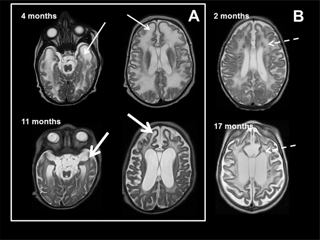
Figure 2. MRI characteristics in AGS.
MR imaging obtained in two patients with AGS at age 4 months (upper row) and 11 months (lower row) of age in box A and the second patient with images at 2 and 17 months (B). The images demonstrate early frontal and temporal lobe swelling (thin white arrow) that gives way to severe frontal and temporal lobe atrophy (thick white arrows), as well as the prominent global atrophy. Note also the T2 dark signal abnormalities presumed to be calcifications present at 2 months and present at 17 months in the second patient (dashed arrows).

