
Fig. 2.
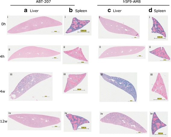
Perl’s Prussian blue staining on liver and spleen sections of rats dosed with ABT-207 or h5F9-AM8. In animals treated with ABT-207, liver iron deposition (a) was visible between week 1 and week 9 and iron reduction (b) in the spleen could be seen within this time frame. In animals treated with h5F9-AM8 (c, d), this observation is more pronounced. Bar represents 3 mm for all panels