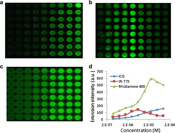
Fig. 1.

Non-linearity of the near-infrared emission intensity in vitro. a–c Representative images of plates incubated with increasing concentrations of indocyanine green (ICG) (a), IR-775 (b), or rhodamine 800 (c). d Quantification of emission intensity. MDCK II cells were seeded in 96-well plates at 8 × 10⁴ cells/well. When achieved monolayers, cells were incubated with the indicated concentrations of the dyes for 1 h, then scanned by a molecular imager. At the studied concentrations, IR-775 and rhodamine 800 exhibited a quenching phenomenon which resulted in decreased signal intensity at higher concentrations. As the dye solution is more saturated, the distance between the fluorochrome molecules decreases and fluorescence resonance energy transfer (FRET) takes place (94). Considering that emission intensity is an indirect measurement of dye’s quantities, this artifact implies lesser concentrations in the cell, while, in fact, the concentrations are higher
