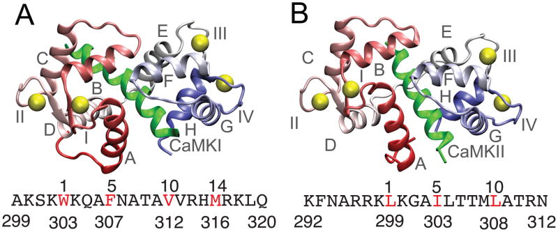
Figure 1.

Native structures of the CaM-CaMBT complexes (A) and (B) show the PDB structures of the CaM-CaMKI (PDB ID: 2L7L) (Gifford et al. 2011) and CaM-CaMKII (PDB ID: 1CDM) (Meador et al. 1993) complexes. Helices and the Ca2+-binding loops of CaM are denoted by A-H and I-IV, respectively in (A) and (B). CaM is colored red (N-terminal domain) and blue (C-terminal domain) and the Ca2+ ions are represented with yellow spheres. The CaMKI and CaMKII peptides are shown in green. The sequence of CaMKI and CaMKII are indicated below (A) and (B), respectively with one letter code. The hydrophobic residues of the CaMBTs from the hydrophobic motifs (1-5-10 and 1-14 for CaMKI and 1-5-10 for CaMKII) in both the sequences are colored in red.
