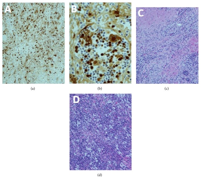
Figure 5.
Immunohistopathology. Section through the left atrial mass shows histiocytes that are immunoreactive to S100 protein immunostaining (a). Emperipolesis is engulfment of lymphocytes and erythrocytes by histiocytes that is considered diagnostic of RDD. Emperipolesis is noted in section from left atrial mass on hematoxylin and eosin-stained sections (b). The histiocytes and lymphoplasmacytic cells infiltrating the myocardium of the left atrium are seen (c). Section from the left atrium also demonstrates fibrosis and large histiocytes in sheets that are accompanied by numerous plasma cells and small mature lymphocytes mass on hematoxylin and eosin-stained section (d).

