
Fig. 2.
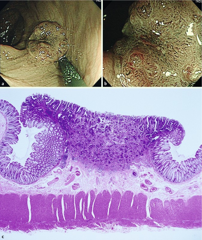
Depressed-type lesion (0 – IIa + IIc), 8 mm in size. The lesion had a deeply depressed area with a non-traumatic tube whose diameter was 2.5 mm, which was classified as NICE 3 (narrow-band imaging international colorectal endoscopic [classification]) with high confidence by NBI-NME (narrow-band imaging with non-magnifying endoscopy) (a) and NBI-ME (narrow-band imaging with magnifying endoscopy) (b). High magnifying endoscopy revealed the interruption of thick vessels. The endoscopists chose to treat with surgery without endoscopic resection. Histology revealed a moderately differentiated tubular adenocarcinoma invading the deep submucosa (c).