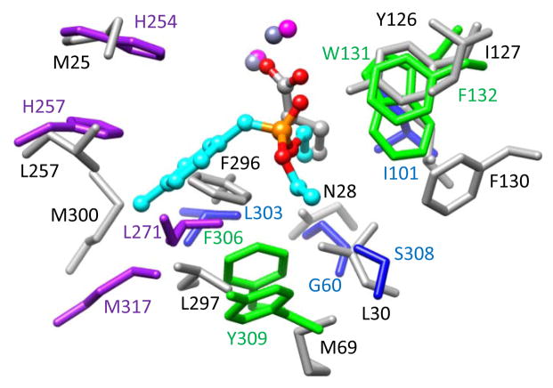
Figure 9.
Superimposition of the residues of the three binding pockets in the active site of PTE (PDB id: 1dpm) and the corresponding residues in Mn-containing structure Pmi1525. For PTE, the large pocket residues are colored in purple, the small pocket residues are colored in blue, and leaving group pocket residues are colored in green. The corresponding residues of Pmi1525 are colored in light grey. The labels of the residues of PTE are colored the same as the residues themselves. The residue labels of Pmi1525 are colored in black.

