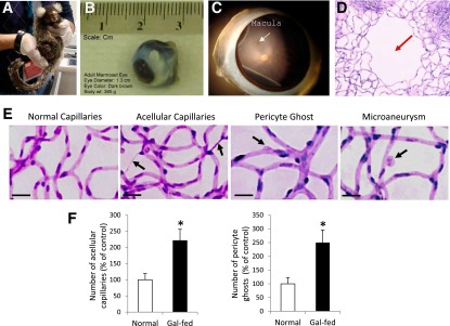
Figure 1.

Marmoset eye, macula, and retinal capillary network surrounding macular area and acellular capillaries, pericyte loss, and microaneurysms in gal-fed marmoset. A: Marmoset is an unusually small primate. B: An adult marmoset eyeball is ∼13 mm in diameter. C: Marmoset macula (white arrow) is present at an ∼2.5 disc diameter distance from the optic disc. D: A prominent foveal avascular zone is located in the macula (red arrow). Note that the relative size and location of macula with respect to the optic disc in the marmoset retina are strikingly similar to those of the human macula. E: Effect of hyperhexosemia on retinal vascular lesions characteristic of DR. Representative images of retinal capillary network show increased number of acellular capillaries and pericyte loss in the retinas of gal-fed marmosets compared with those of control marmosets. Incipient microaneurysms were detected in the gal-fed marmosets. Magnification bar: 20 μmol/L. F: Graphical illustrations showing significant increase in the number of acellular capillaries (left) and pericyte ghosts (right) in the retinas of gal-fed marmosets compared with those of control marmosets. *P < 0.05.
