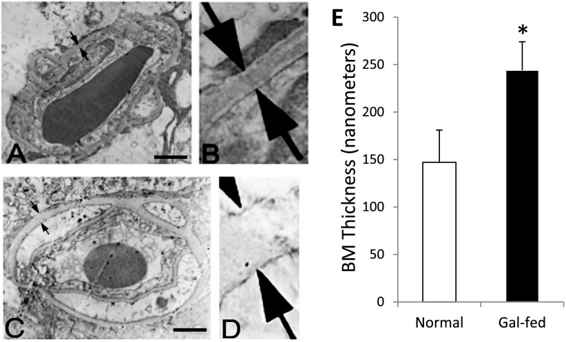
Figure 2.
Capillary BM thickness in retinas of marmoset. BM thickness (arrows) in transverse sections of a retinal capillary from normal marmoset (A). B: Corresponding enlarged view. C: Gal-fed marmoset. D: Corresponding enlarged view. Magnification bar: 1 μmol/L. E: Graphical illustration of capillary BM thickness in retinas of normal and gal-fed marmoset. *P < 0.02.

