
Figure 3.
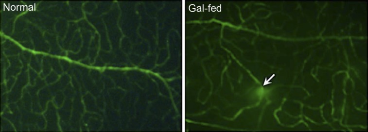
Retinal vascular permeability in gal-fed marmoset. Representative areas showing FITC fluorescence in vessels of whole-mount retinas. Areas of intense FITC leakage from extravasation were observed in the retinas of gal-fed marmosets compared with those of control normoglycemic marmosets. In the gal-fed marmoset retinas, some vessels showed vascular leakage at specific points of the vessel (arrow).