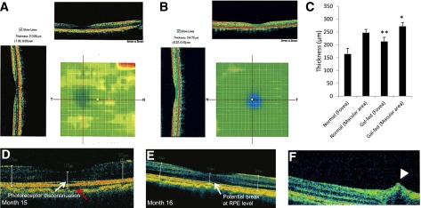
Figure 4.

Representative retinal OCT scans of a gal-fed marmoset and a normal marmoset and pathological changes in the retinas of gal-fed marmosets. Significant thickening of the foveal and the juxtafoveal area, indicative of intraretinal fluid accumulation, was observed in retinas of gal-fed marmosets. Color heat images show increased foveal thickness in retinas of a gal-fed marmoset (A) compared with the foveal thickness of a normal marmoset (B). C: Graphical illustration showing macular and foveal thickness in retinas of control and galactose-fed marmosets. *P < 0.03, **P < 0.02. D–F: Representative images of the macular area showing alterations in RPE, photoreceptor layer, and an incipient cystoid retinal edema in gal-fed marmosets. D: Fluid accumulation in the choroid (red arrow) and discontinuation at the photoreceptor layer (white arrow) representing early stages of edematous retina were observed in a gal-fed marmoset. E: Retinal thickening was accompanied by changes in RPE and retinal photoreceptor layer starting at the fifteenth month of galactose feeding. F: An OCT scan of the central retina showing a hyporeflective space just above the RPE level indicative of intraretinal fluid accumulation (arrowhead).
