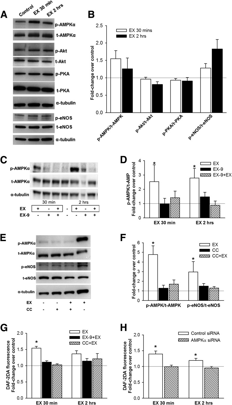
Figure 5.
The effects of exenatide in vitro in human endothelial cells. A and B: Phosphorylation of AMPKα (Thr172), PKA (Thr197), Akt kinase (Ser473), and eNOS (Ser1177) in HAECs after treatment with 10 nmol/L exendin-4 (EX) for 30 min and 2 h (A: representative Western blot; B: densitometry analysis [means ± SE]; n = 6). C and D: AMPKα phosphorylation in HAECs after EX (30 min and 2 h) with or without pretreatment for 30 min with 1 μmol/L GLP-1R inhibitor EX-9 (C: representative Western blot; D: densitometry analysis; n = 7–8). E and F: AMPKα and eNOS phosphorylation in HAECs after EX (2 h) with or without 1-h pretreatment with 5 mol/L of AMPKα inhibitor CC (E: representative Western blot; F: densitometry analysis; n = 8–9). The effect of EX on NO production (by DAF-2DA fluorescence) in HAECs with or without pretreatment with EX-9 or CC (n = 4–7) (G) and in HUVECs with knocked-down AMPKα gene expression (siRNA, n = 6) (H). Phosphorylated bands were normalized to total bands and α-tubulin. Control, untreated cells. Data are means ± SE. *P < 0.05 vs. control.

