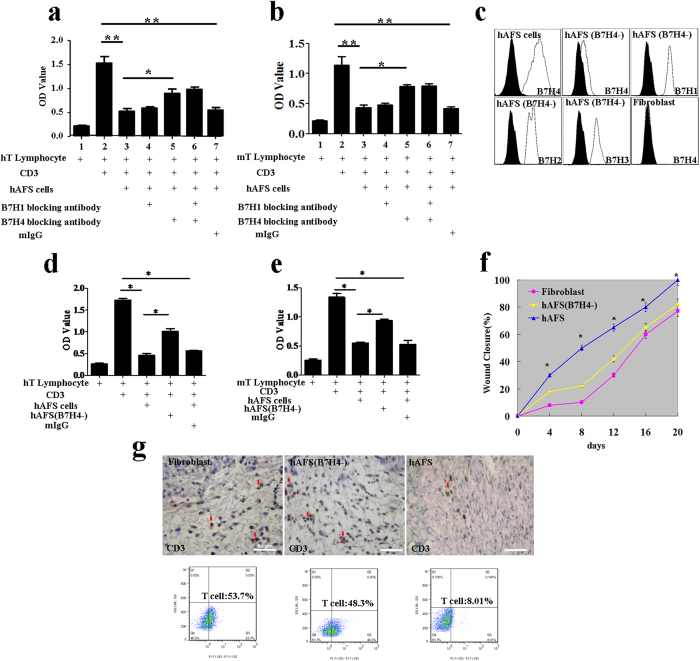
Figure 6. The role of the B7H4 molecule in epidermal regeneration.
(a) Analysis of the inhibitory effect of hAFS cells on human lymphocytes and the effect of the B7 family blocking antibody on hAFS inhibitory ability. The data are presented as the mean ± SD of three independent experiments. Analysis was performed with GraphPad Prism; *p < 0.05, **p < 0.01. (b) Analysis of the inhibitory effect of hAFS cells on mouse lymphocytes. The effect of the B7 family blocking antibody on hAFS cells’ inhibitory ability. The data are presented as the mean ± SD of three independent experiments. Analysis was performed with GraphPad Prism; *p < 0.05; **p < 0.01. (c) hAFS cells, fibroblasts and B7H4-downregulated hAFS cells were analysed by flow cytometry after staining with PE-conjugated control isotype IgG (black peaks) or antibodies against the cell surface proteins B7H4. B7H4-downregulated hAFS cells were also analysed by flow cytometry after staining with PE-conjugated control isotype IgG (black peaks) or antibodies against the cell surface proteins B7H1, B7H2 and B7H3. (d) Analysis of the inhibitory effect of B7H4-downregulated hAFS cells on human lymphocytes. (e) Analysis of the inhibitory effect of B7H4-downregulated hAFS cells on mouse lymphocytes. (f) Measurement of wound closure in the B7H4-downregulated hAFS, fibroblast, and hAFS cell groups in BALB/c mice. Analysis of variance (ANOVA) of wounds measured using the UTHSCSA ImageTool; *p < 0.01. (g) Infiltration of CD3+ T cells (red arrow) into the granulation tissue of BALB/c mice in the fibroblast, B7H4-downregulated hAFS and hAFS groups at day 4. When compared with the hAFS group, more infiltrated CD3+ T cells are visible in the fibroblast and B7H4-downregulated hAFS groups.

