
Figure 2.
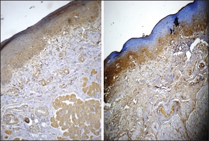
Grade II oral submucous fibrosis stained with antitransforming growth factor-beta1 (TGF-β1) (left side picture) and anti-TGF-β2 (right side picture). Note restriction of TGF-β2 expression to submucosa, especially subepithelial (original magnification, ×10)