
Fig. 2.
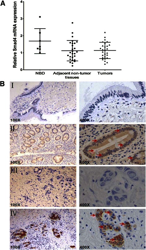
The expression levels of Smad4 in the primary EHCC and NBD specimens. a The mRNA expression level of Smad4 in the primary EHCC, the adjacent non-tumor tissues and NBD specimens determined by qRT-PCR. U6 was used as the internal control. b Immunohistochemical staining of Smad4 protein expression in primary EHCC and NBD samples. The arrows indicated nuclear staining of Smad4 in both NBD and EHCC samples. (I) Low Smad4 protein expression in a NBD. (II) High Smad4 protein expression in a NBD. (III) Reduced Smad4 protein expression in a primary EHCC. (IV) High Smad4 protein expression in a primary EHCC. Original magnification, 100× and 400× respectively for each slide