Figure 1.

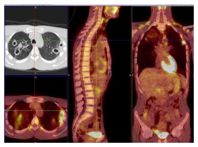
M. kansasii infection, cavitary lesions in the right upper lobe and in the apical segment of the right lower lobe and left upper lobe, SUV max 5.36. Related to patient 3 (PET/CT. Courtesy “V. Monaldi” Hospital).

M. kansasii infection, cavitary lesions in the right upper lobe and in the apical segment of the right lower lobe and left upper lobe, SUV max 5.36. Related to patient 3 (PET/CT. Courtesy “V. Monaldi” Hospital).