
Figure 4. Absent KIAA0586 protein in patient fibroblasts.
Immunoblot analysis of KIAA0586 in fibroblasts from family MTI-103 and MTI-233. Lysates from RPE1 cells transfected with scrambled or KIAA0586 siRNA were used as control. M, unaffected carrier (mother); A, affected child. RPE1, retinal pigment epithelial-1 cell line. Figure 4—figure supplement 1A represents an expression analysis of the KIAA0586 gene.
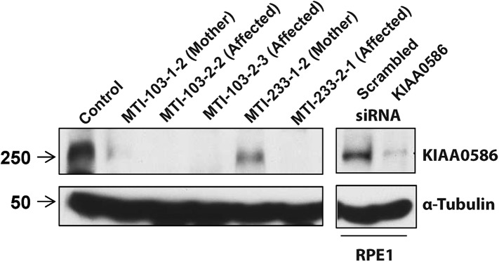
Figure 4—figure supplement 1. Expression analysis of the KIAA0586 gene.
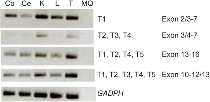
RT-PCR analysis showing differential expression levels of the KIAA0586 transcripts amongst various ciliated and non-ciliated tissues was observed. Co, colon; Ce, cerebellum; K, kidney; L, liver; MQ, MilliQ; T, testis; T1-T5 transcript number corresponding to Figure 2A.