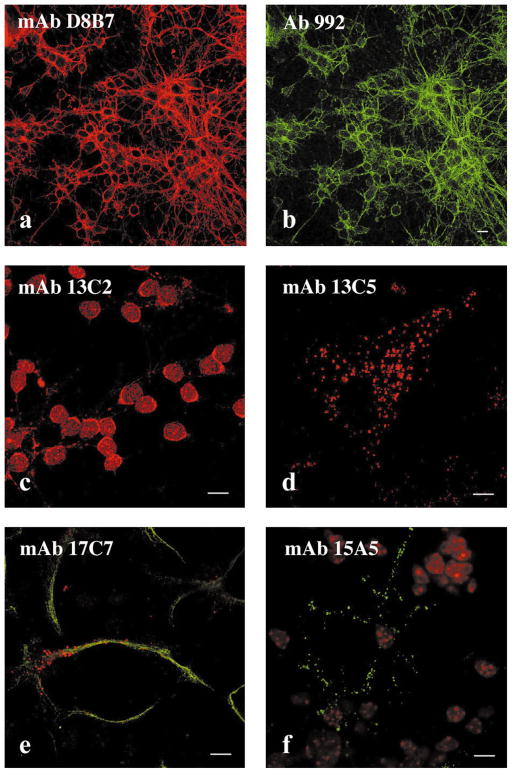
Fig. 3.
Immunostaining pattern of monoclonal antibodies to spectrin SH3 domains. Mouse cerebellar cultures (a–c and e and f) and NIH 3T3 cells (d) were stained with monoclonal antibodies to spectrin SH3 domain. A culture enriched for granule neurons costained with mAb D8B7 to αII SH3 domain (a, red) and with the polyclonal antibody 992 to axonal spectrin [17] (Chemicon, Temecula, CA) (b, green), or stained with mAb 13C2 (c) (red). A culture of cerebellar cells enriched for astrocytic cells costained with mAb 17C7 (e, red) and with the polyclonal antibody to GFAP (Sigma, St. Louis, MO) (f, green). An NIH 3T3 fibroblast stained with mAb 15A5 (d) (green), nuclear autofluorescence (red). Antibodies 17C7, 13C2, and 15A5 produce very similar pattern in NIH 3T3 cells to that observed with mAb 13C5. Note coincident pattern in (a) and (b), indicating colocalization of antibody staining. Bar, 10 μm.

