Image 5.
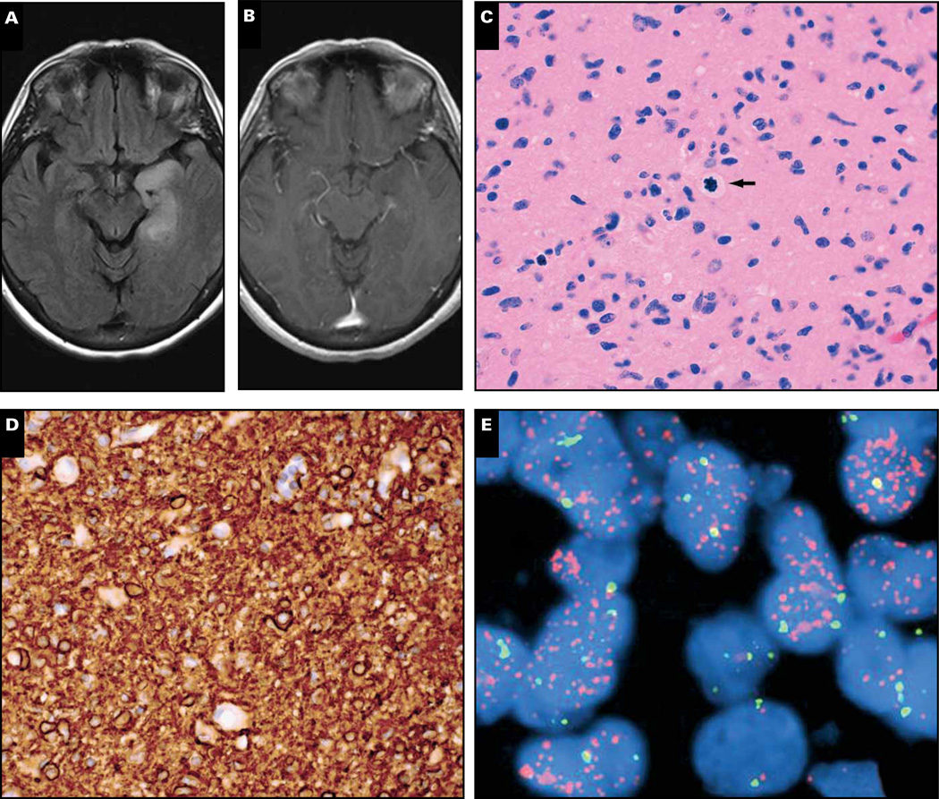
Case 7. A 61-year-old woman with left-sided seizures was found to have a left temporal lesion that showed a T2 signal but no significant contrast enhancement on T1 magnetic resonance imaging (MRI) (A, B). Histologic examination of the resection specimen showed an infiltrating glial tumor with grade III histology, including angulated atypical nuclei and readily identified mitoses (C, arrow); no microvascular proliferation or necrosis was seen. The tumor showed strong, diffuse immunohistochemical expression of EGFR (D), and EGFR was amplified (E). The tumor was negative for IDH1/2 mutations via both immunohistochemistry and polymerase chain reaction (not shown). Orange signals (E), EGFR; green signals, chromosome 7 centromeric enumeration probe.

