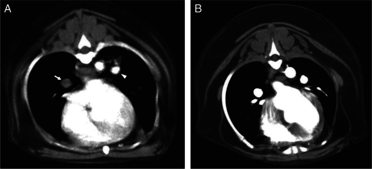
FIG 1.

CT pulmonary angiography (CTPA) from two dogs with immune-mediated haemolytic anaemia. (A) Positive CTPA study diagnostic for PTE. Intraluminal filling defects can be clearly seen in both the right (arrow) and left (arrowhead) main pulmonary arteries. The filling defect in the left pulmonary artery is only partial at this level. (B) Negative CTPA study which rules out PTE in this patient. There is normal opacification of both left at right pulmonary arteries by contrast at this level. No aortic filling defects were noted in this study
