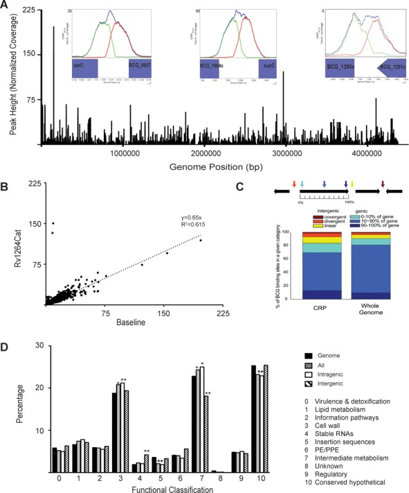
Figure 1.

Binding of the CRPBCGin vivo. (A) Distribution of CRPBCG binding across the genome is evenly dispersed. Within insets are select, known binding sites and the correlating ChIP-Seq binding peaks. (B) Side-by-side plot of one repeat of untreated cells (baseline) against peaks for the Rv1264Cat. (C) Distribution of the binding sites within the various regions of genes. (D) Functional classification of the ChIP-Seq binding peaks within various categories including only intergenic sites, intragenic sites and all sites plotted together. *P at least <0.05 relative to the genome using a hypergeometic calculation with a correction factor of 11, **P < 0.005.
