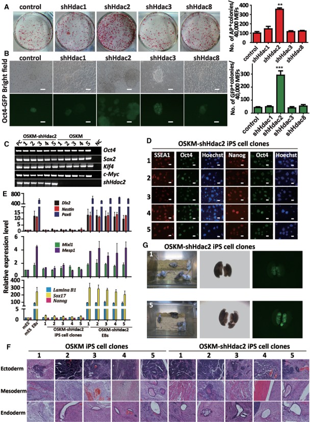
Figure 1.

Knockdown of Hdac2 improves the generation of OSKM-iPS cells. (A) AP staining (on reprogramming day 8) of colonies from OG-MEFs transduced with OSKM in combination with control shRNA, shHdac1, shHdac2, shHdac3 or shHdac8. AP-positive colony numbers are shown on the right of the figure. (B) The morphology of typical Oct4-GFP iPS cell colonies on reprogramming day 12 in combination with shHdac1, shHdac2, shHdac3 or shHdac8, respectively. GFP-positive colony numbers are shown on the right. Scale bar, 100 μm. (C) Genomic PCR analyses showed that the OSKM-shHdac2-derived iPS cell clones (1–5) were infected with OSKM and shHdac2 simultaneously. OG-MEFs were used as a negative control (NC) and OG-MEFs infected with the OSKM viruses or shHdac2 were used as positive controls (PC). (D) Immunostaining for pluripotency markers (Oct4, Nanog and SSEA-1) in the OSKM-shHdac2-derived iPS cell clones. Scale bar, 100 μm. (E) qRT-PCR analyses of markers for all three germ layers (Dlx2, Nestin and Pax6 for ectoderm, Mixl1 and Mesp1 for mesoderm, and Lamina B1 and Sox17 for endoderm) and a pluripotency factor (Nanog) in mES cells, mES EBs, OSKM-shHdac2 iPS cell clones and OSKM-shHdac2 EBs. The expression levels were normalized to Gapdh. (F) OSKM-shHdac2-derived iPS cell clones induced teratomas containing all three embryonic germ layers. Representative images of HE staining for neural tissue (ectoderm), skeletal muscle (mesoderm) and epithelial tissue (endoderm) are shown. (G) Two-week-old chimeric mice derived from OSKM-shHdac2-1and OSKM-shHdac2-5 iPS cell clones (C57BL/6 background). Oct4-GFP positive cells derived from OSKM-shHdac2-1 or OSKM-shHdac2-5 iPS cell clone are present in the genital ridge of a female embryo at 13.5 day postcoitum (dpc). The data in a, b and e represent the means ± S. E. M. of three independent experiments. **P < 0.01, ***P < 0.001 (two-tailed Student's t-test).
