
Figure 4.
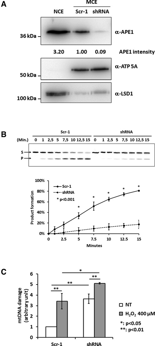
Loss of APE1 expression determines increased levels of mtDNA damage. (A) Western blot analysis of mitochondrial fractions (MCE) from control (Scr-1, lane 2) and APE1-shRNA stable clones (lane 3). APE1-shRNA cells show APE1 reduction up to 90% with respect to the Scr-1 cells. ATP 5A and LSD1 were used as mitochondrial and nuclear markers, respectively. A nuclear extract (NCE, lane 1) from HeLa cells was used as control to exclude nuclear/mitochondrial cross contamination. (B) Endonuclease activity analysis of mitochondrial extracts from Scr-1 and shRNA cells. The conversion of the fluorescent tetrahydrofuran-containing oligonucleotide substrate (S) to the shorter incised product (P) was evaluated for the reported times on a denaturing 20% (wt/vol) polyacrylamide gel. A representative image (top) and average values of incision percentage ±SD of three independent experiments (bottom) are shown. Endonuclease activity is almost abolished upon loss of APE1 expression. (C) mtDNA damage analysis of Scr-1 and shRNA clones under basal conditions and after H2O2 treatment (400 μM). Levels of mtDNA damage are increased in cells lacking APE1 both under basal and oxidative stress conditions.