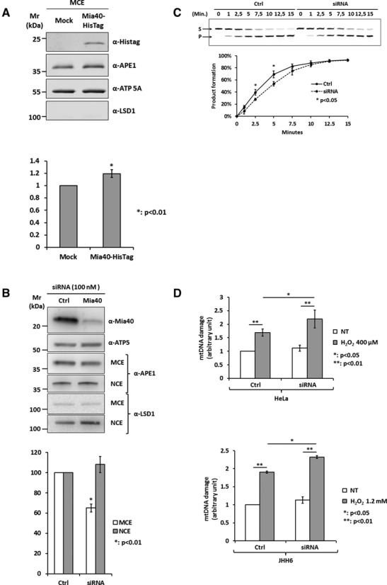
Figure 5.

Loss of Mia40 expression reduces mitochondrial APE1 content affecting mtDNA stability. (A) Western blot analysis (top) of mitochondrial extracts from HeLa cells overexpressing Mia40-HisTag. HeLa cells were transiently transfected with pCMV 5.1 empty vector (Mock) or codifying for Mia40-HisTag protein. Densitometric analysis of mitochondrial APE1 levels (bottom) highlights a statistically significant increase of APE1 content into the mitochondrial fraction as a consequence of Mia40 overexpression. Data are the mean ± SD of three independent experiments. (B) Western blot analysis of nuclear (NCE) and mitochondrial (MCE) HeLa cell extracts of Mia40 siRNA cells (top). Densitometric analysis of mitochondrial and nuclear APE1 levels (bottom) highlights a statistically significant reduction of APE1 content into the mitochondrial fraction as a consequence of Mia40 loss of expression. Data are the mean ± SD of five independent experiments. (C) Endonuclease activity analysis of mitochondrial fractions from scramble (Ctrl) and Mia40-siRNA-treated cells (siRNA). Data analysis reported in the diagram (bottom) shows that endonuclease activity is reduced after Mia40 silencing. (D) mtDNA damage analysis from HeLa cells (top) and JHH6 hepatic cell line (bottom) after Mia40 loss of expression under basal conditions and after H2O2 treatment (HeLa: 400 μM; JHH6: 1.2 mM). Silencing of Mia40 leads to an increase of mtDNA damage under oxidative stress conditions.
