
Figure 5.
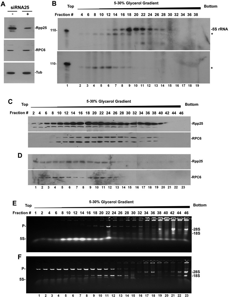
Knockdown of Rpp25 prevents formation of initiation complexes. (A) Western blot analysis of Rpp25, RPC6 and α-Tubulin in HeLa cells transfected for 48 h with a minigene expressing a siRNA directed against Rpp25 or with an empty plasmid (24). (B) Transcription of 5S rRNA gene by initiation complexes purified by velocity sedimentation in large-scale 5–30% glycerol gradients (see Materials and Methods section). Initiation complexes were derived from whole extracts of mock-transfected cells (with plasmid; upper panel) or cells transfected with a minigene expressing a siRNA against Rpp25 (lower panel). The leftmost lane shows a 110-nt RNA size marker. Asterisks point to labeled non-specific RNAs. (C and D) Western blot analysis of Rpp25 and RPC6 in fractions described in (B). Based on the exposure times of the blots, the level of Rpp25 in panel (D) was reduced by ∼90%, when compared with that seen in (C). (E and F) Detection of 5S rRNA, 18S rRNA and 28S rRNA in the indicated fractions described in (B) was performed as in Figure 3E. The level of 5S rRNA was low (lower panel) due to inhibition of transcription of endogenous 5S rRNA genes in transfected cells deficient in Rpp25.