
Figure 4.
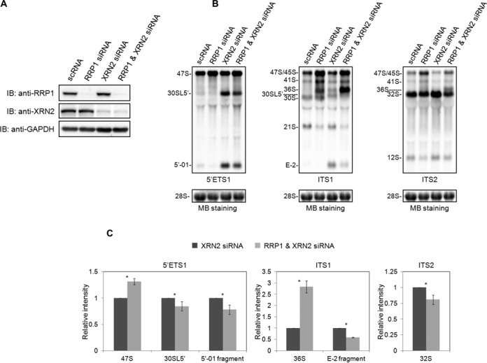
RRP1 accelerates the site 2 cleavage of 47S/45S pre-rRNA. (A) Total protein from HeLa cells (20 μg/lane) treated with scRNA, RRP1 siRNA, XRN2 siRNA, or RRP1 and XRN2 siRNA was analyzed by immunoblotting (IB) with the antibodies indicated at the left. (B) Pre-rRNA intermediates isolated from HeLa cells treated as in (A) were detected by northern blot analysis using 5′ ETS1, ITS1 and ITS2 probes as indicated at the bottom of each staining set (top). 28S rRNA was visualized by methylene blue staining (MB staining) as a loading control (bottom). The pre-rRNA species are assigned at the left of each staining set. (C) The intensities of the 47S, 30SL5′ pre-rRNA and 5′-01 fragment detected by 5′ ETS1, of the 36S pre-rRNA and E-2 fragment detected by ITS1, and of the 32S pre-rRNA detected by ITS2 were quantified, first normalized to the amount of 28S rRNA and then graphed relative to the amount of the corresponding band from XRN2 siRNA-treated cells. The values are averages (± SD) of three independent experiments. *, P < 0.05.