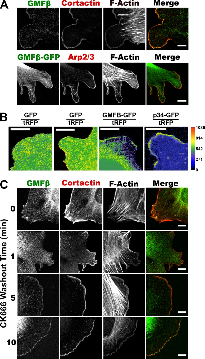
Figure 1.
GMFβ localizes to the leading edge of fibroblasts. (A) GMFβ localization by immunofluorescence (IF; top) and in cells expressing GMFβ-GFP (bottom). Arp2/3 or cortactin IF marks leading edge. (B) Ratio of either soluble GFP, GMFβ-GFP, or p34-GFP to soluble RFP. The legend to the right represents pixel intensity. (C) IF for GMFβ and cortactin of cells treated with CK-666 (150 µM) to ablate lamellipodia, followed by washout for given times to allow lamellipodia regrowth. Bars, 10 µm

