Figure 4.
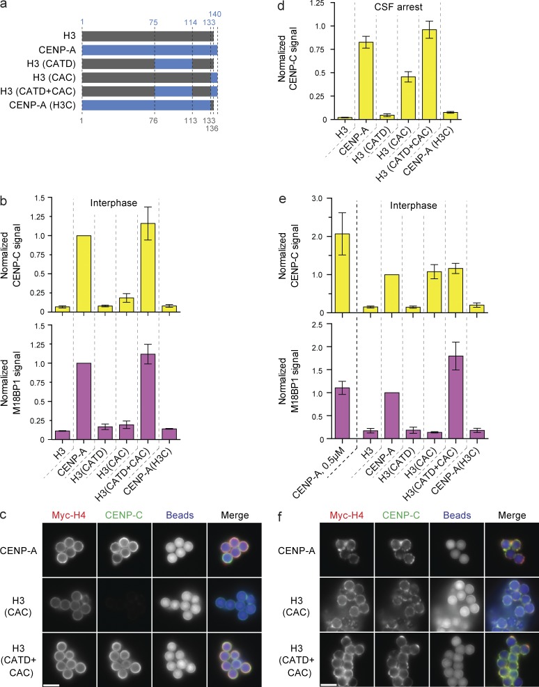
CAC-mediated CENP-C recruitment is dependent on high nucleosome density. (a) Schematic of the histone chimeras used and the amino acid residues of human CENP-A (blue) and histone H3 (gray), respectively. (b) CENP-C (top) and M18BP1 (bottom) levels on low saturation chimeric nucleosome arrays in interphase. Signals are compared with the amount of recruitment to wild-type CENP-A beads and internally normalized in each sample to the levels of Myc-H4 on the beads. Bars in all panels represent the means ± SEM; n = 3. (c) Fluorescent images of CENP-C recruitment to low-saturation CENP-A, H3(CAC), and H3(CATD+CAC) chromatin arrays. The Myc-H4, CENP-C, and bead autofluorescence signals are shown. (d) CENP-C levels on CENP-A/H3 chimeric nucleosome arrays reconstituted at 0.5 µM nucleosome concentration in CSF-arrested extract. Signals are normalized as in b. (e) CENP-C (top) and M18BP1 (bottom) levels on high-saturation chimeric nucleosome arrays. Signals are normalized as in b. The signal on low-saturation CENP-A arrays (CENP-A, 0.5 µM) are shown for comparison. (f) Fluorescence images of CENP-C levels on high-saturation CENP-A, H3(CAC), and H3(CATD+CAC) chromatin arrays. Images labeled as in c. Bars, 5 µm.

