Figure 2.

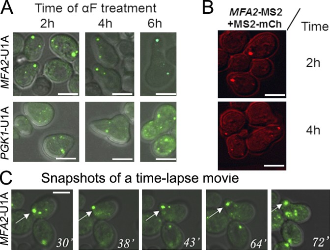
MFA2 mRNA granules, but not PGK1 mRNA granules, are preferentially localized to shmoo tips. (A) Microscopic analysis of the distribution of low- and high-intensity MFA2 and PGK1 mRNA granules during shmoo growth. The WT strain coexpressing U1A-GFP and either MFA2-U1A (ySA20) or PGK1-U1A (ySA32) was treated with αF for the indicated time. n = 200–250 from three independent experiments. (B) Shmoo localization of MFA2 mRNA granules was observed using an MS2 labeling system, instead of the U1A system. The WT strain (ySA24) expressing MFA2-MS2 (from pMC475) and MS2-mCh (from pMC522) was grown in selective medium until mid–log phase and then treated with αF. At the indicated time after αF addition, cells were inspected under confocal microscope. (C) Snapshots from time-lapse analysis of MFA2 mRNA-containing granules during shmoo formation in response to αF treatment (Video 1). The arrows point at a high-intensity MFA2 mRNA granule that was located in the shmoo tip during the growth of the shmoo (Video 1). Bars, 5 µm.
