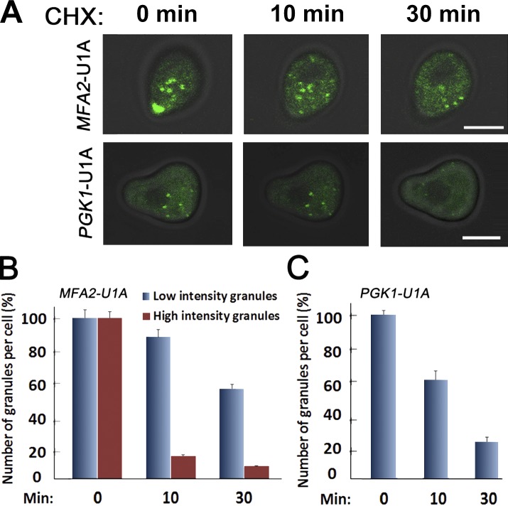
Figure 7.
The large granules of MFA2 mRNA dissociate rapidly in response to CHX. (A) Cells expressing MFA2-U1A (ySA20) and PGK1-UA1 mRNA (ySA32) detection system were treated with αF for 2 h. 100 µg/ml cycloheximide (CHX) was then added, and cells were inspected microscopically 10 or 30 min later (top). Bars, 5 µm. (B and C) The percentage of high- and low-intensity MFA2 or PGK1 mRNAs granules was determined at the indicated time points after CHX addition. The percentage is plotted relative to t = 0 (before CHX addition), which was arbitrarily defined as 100%. Error bars represent the standard deviation of three independent experiments.

