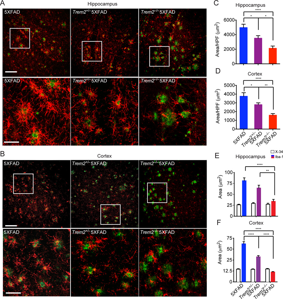
Figure 3. TREM2 deficiency leads to reduced microgliosis in 5XFAD mice.
Microgliosis in 8.5 month-old Trem2−/−5XFAD, Trem2+/−5XFAD and 5XFAD mice. (A, B) Matching coronal sections were stained with Iba-1 (red) for microglia and X-34 (green) for amyloid plaques. Representative Z-stack images with maximum projection are shown. (C–D) Quantification of total Iba-1 reactivity per high power field (HPF) in hippocampi and cortices. (E, F) Quantification of microgliosis associated with plaques of similar sizes in hippocampi and cortices. Original magnification 20× (A, B, upper panels), 40× (A, B, lower panels); Scale bar= 10µm (A, B, upper panels), 50 µm (A, B, lower panels). *p<0.05, **p<0.01, ****p<0.0001, one-way ANOVA. Data represent analyses of a total of 8–10 5XFAD, 8–12 Trem2+/− 5XFAD mice, and 8–16 Trem2−/−5XFAD mice. Bars represent mean±SEM. See also Figure S3 and Movie S1–3.

