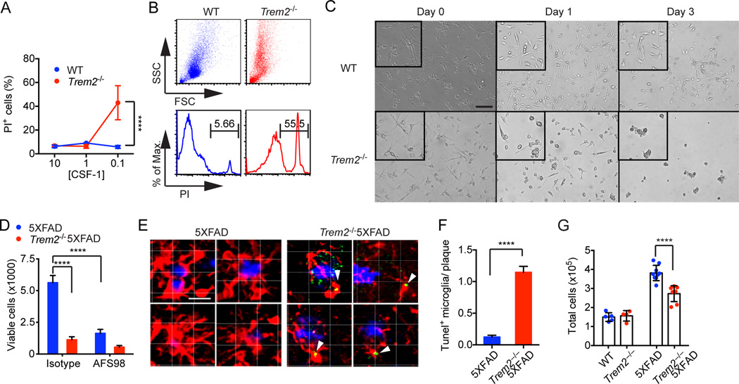
Figure 5. TREM2 promotes microglia survival ex vivo and in vivo.
(A–C) Adult primary microglia were cultured with various concentration of CSF-1-containing L-cell medium (LCM). Viability of microglia by PI staining (A–B) and morphology (C) were assessed on day 3. (D) Microglia were purified ex vivo from 5XFAD mice and cultured in 0.1% LCM with or without CSF-1R blocking antibody AFS98. Viability was determined on day 5. (E, F) Apoptosis of plaque-associated microglia cells (Iba-1, red) in 5XFAD and Trem2−/−5XFAD mice was determined by TUNEL staining (green). Plaques were identified by X-34 (blue). Representative single-stack images of 5XFAD and Trem2−/−5XFAD microglia (E) and summary of frequencies of TUNEL+ microglia associated with plaques (F) are shown. Original magnification: 20×; scale bar= 10µm (C), 15µm (E). (G) Total numbers of live microglia cells in cortices and hippocampi of 5XFAD, Trem2−/− 5XFAD, Trem2−/− and WT mice. ****p<0.0001, two-way ANOVA (A, D, G), student's t-test (F). Data represent a total of three independent experiments (A–D) and a total of 5–8 mice per group (E–G). Bars represent mean±SEM. See also Figure S5 and Movie S4.

