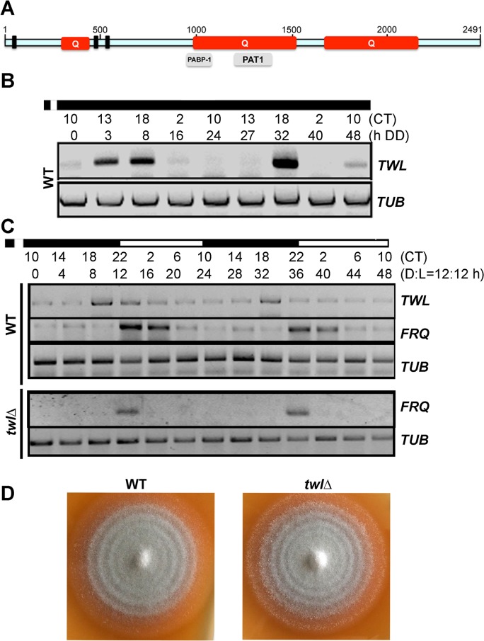
Fig 1. Twl is a novel circadian protein in M. oryzae.
(A) Schematic representation of the domains in M. oryzae Twl protein sequence. Q, glutamine-rich region. Three short bars represent P-loop pattern [AG]-x(4)-[AG]-[KR]-[ST], identified by ScanProsite (http://prosite.expasy.org/scanprosite/). Grey bars in lower panel are partial domains predicted by NCBI. The schematic is drawn to scale based on the amino acid sequence. (B) Circadian rhythm of TWL in the wild type. WT mycelia were grown in the dark for 5 d, and then briefly (30 min) exposed to light, before a 48 h (two circadian cycles) growth in constant dark. Total RNA was extracted at the indicated time points for semi-quantitative reverse transcriptase PCR. CT, Circadian Time. CT12 corresponds to the time point immediately following light exposure. (C) Circadian rhythm of TWL and FRQ in the entrained wild type or the twlΔ mutant. WT or the twlΔ mycelia were grown in liquid Complete Medium (CM) in dark for 3 d, and then briefly (30 min) exposed to light, before a 48 h (two Dark/Light = 12h /12 h cycles) growth. Total RNA was extracted at four-hour intervals for semi-quantitative reverse transcriptase PCR. CT, Circadian Time. CT12 corresponds to the time point immediately following light exposure. (D) WT and the twlΔ mycelial plugs were inoculated on Prune Agar (PA) medium and kept in constant dark for three days, and then shifted to dark/light (12h /12 h) cycles. The conidial banding patterns in the WT and the twlΔ colonies were imaged after 4 dark/light cycles.

