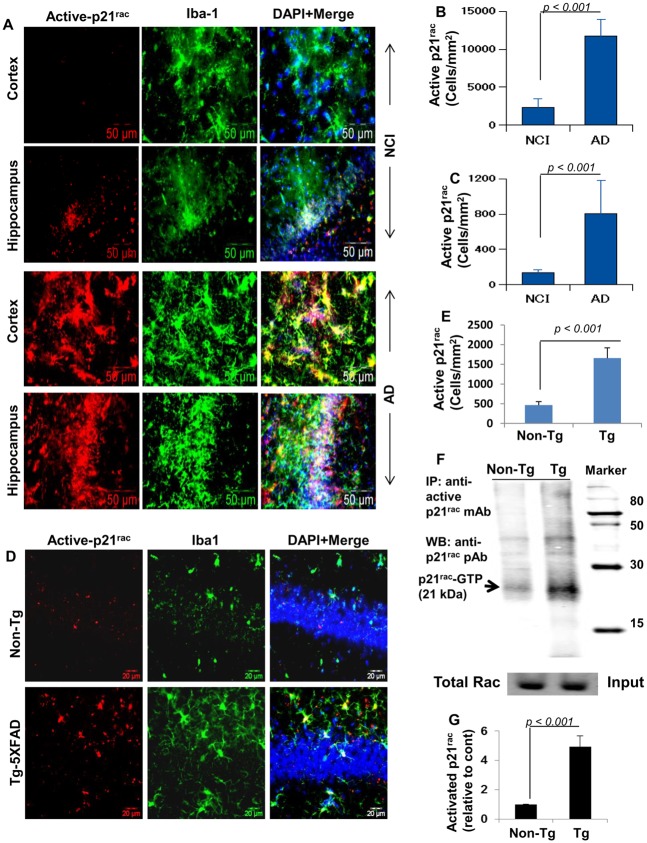
Fig 4. Monitoring activation of p21rac in vivo in the CNS of AD subjects and 5XFAD transgenic mice.
(A) Cortical and hippocampal sections of cases with no cognitive impairment (NCI) and AD were double-labeled with Iba-1 (microglia) and active p21rac (GTP-bound p21rac). Results represent analysis of two sections from each of four different brains. Active p21rac-positive cells were counted in two sections (two images per slide) of each of four different brains per group in an Olympus IX81 fluorescence microscope using the MicroSuite imaging software (B, cortex; C, hippocampus). D) Six month old 5XFAD Tg mice and background- and age-matched non-Tg mice were perfused with PBS-paraformaldehyde followed by double-label immunofluorescence analysis of hippocampal sections for active p21rac and Iba1. Results represent analysis of three sections of each of five mice per group. E) Active p21rac-positive cells were counted in three sections (two images per slide) of each of five different brains per group. F) To further monitor the activation of p21rac, hippocampal homogenates of non-Tg and Tg mice were immunoprecipitated with antibodies against active p21rac followed by Western blot with antibodies against total p21rac. Total p21rac in input was run as experimental control. G) Bands (21kDa) were scanned and presented as relative to non-Tg. Results represent analysis of four mice per group.

