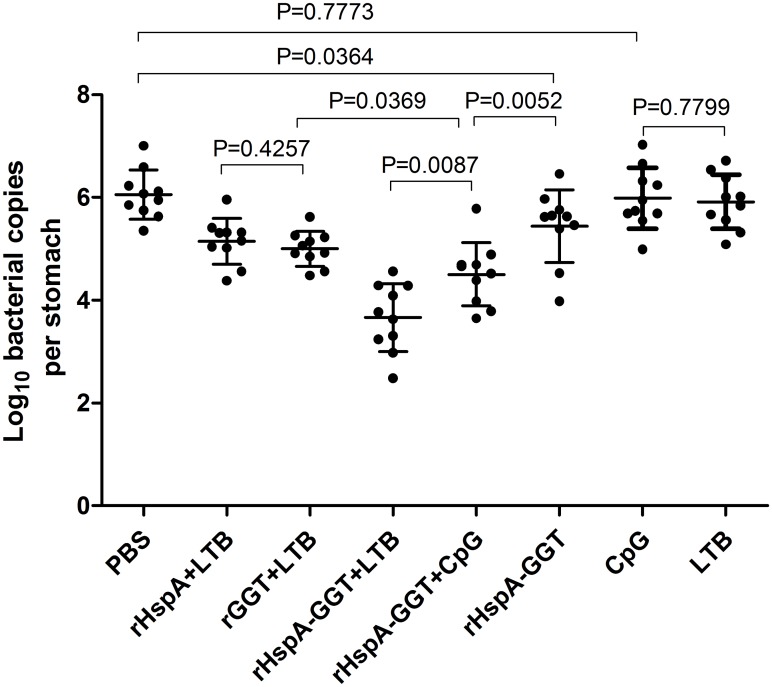
Fig 2. H. pylori colonization in mouse stomachs after immunization.
BALB/c mice (n = 10) were immunized intranasally on days 0, 14 and 21 with 30 μg antigen, with or without 10μg adjuvant, as indicated in Table 1. The same volume of PBS was used as a negative control. Three weeks after the final vaccination boost, mice were orally challenged four times with H. pylori B6. The level of gastric H. pylori colonization was determined by real-time quantitative PCR four weeks post challenge for each mouse. Data are expressed as mean ± S.D. Significant differences between indicated groups are presented as P values.

