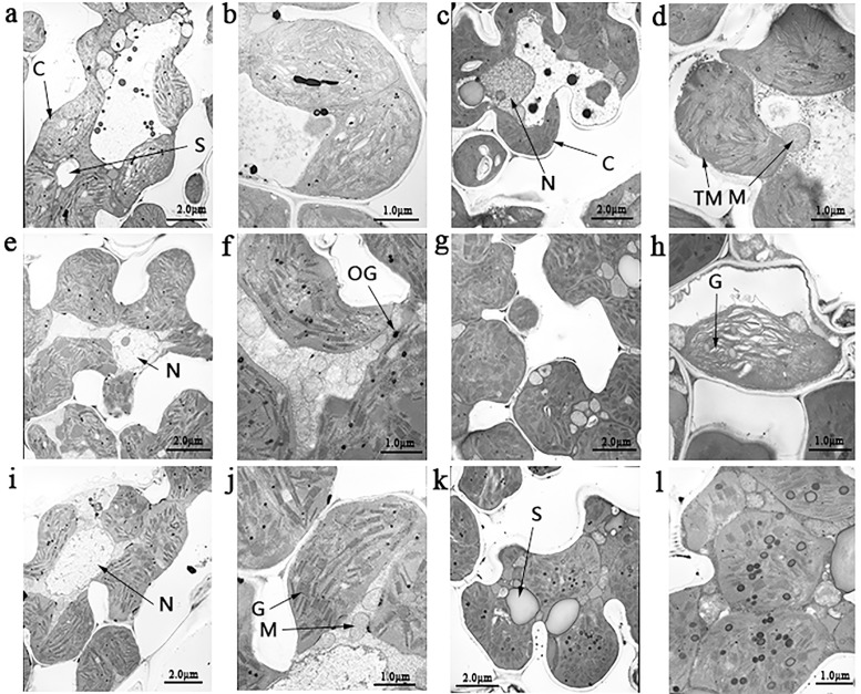
Fig 2. Ultrastructure of chloroplasts in the mesophyll cells of mutant plants.
a, shown are transmission electron microscopy images of chloroplast of the wild type (WT) in 5000 times in tillering stage; b, enlarged in 20000 times for WT in tillering stage; C, 5000 times for WT in jointing stage; d, 20000 times for WT in jointing stage; e, st1-2 at 5000 times in tillering stage; f, st1-2 at 20000 times in tillering stage; g, st1-2 at 5000 times in jointing stage; h, st1-2 at 20000 times in jointing stage; i, st1-3 at 5000 times in tillering stage; j, st1-3 at 20000 times in tillering stage; k, st1-3 at 5000 times in jointing stage; l, st1-3 at 20000 times in jointing stage; C, chloroplast; F, fat; G, grana; M, mitochondria; N, nucleus; OG, osmiophilic plastoglobuli; S, starch granule; TM, thylakoid membranes.

