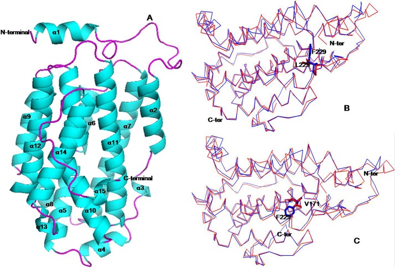
Fig 6. The putative tertiary structure of small subunit of ribonucleotide reductase 1.
(A) Cartoon representation of RNRS1 molecular structure. (B) Comparison of the overall structure between RNRS1 (red) and st1-2 (Val171 mutation, blue) and (C) st1-3 (Leu229 mutation, blue). All figures were prepared using PyMOL. The mutation sites were represented by sticks.

