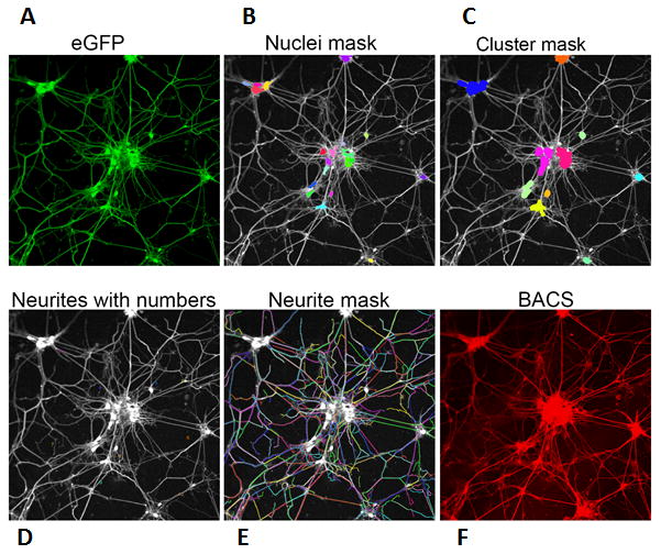
Figure 2. Steps involved in the image analyzing algorithm for detecting endogenous SNAP-25 specifically in motor neurons.

High-content imaging (HCI) can measure the effects of BoNTs and/or small molecules on neuronal morphological changes. Mouse embryonic stem cells (HBG3 line), in which eGFP expression is driven by a motor neuron specific promoter (Hb9), were differentiated into motor neurons. (A) eGFP signal was utilized to identify motor neurons. (B) The fluorescent signal from the eGFP channel was used to detect and mask nuclei and neurite outgrowth. Capella (PerkinElmer)-based nuclei and neurite detection algorithms were used to identify nuclei and neurite outgrowth, respectively. (C-E) A cluster detection module was inserted in the imaging analysis pipeline to detect nuclei clusters using nucleus masks. (F) Without the toxin exposure, BACS antibody signal exhibits total SNAP-25 in eGFP+ cells.
